Fact Sheet Dog Breed Pug
Species: Dog
Breed: Pug
QUEN-Fact Sheet Nr. 29-EN
Status: 30.10.2024
Species: Dog
Breed: Pug
QUEN-Fact Sheet Nr. 29-EN
Status: 30.10.2024
1. Description of the animals
External appearance and critical features required by the standard:
According to the breed standard, the Pug is a „multum in parvo“ (a lot (muscles) in little (space)). It is characterised by its square and chunky build, which is also referred to as compact. A slight rolling of the hindquarters characterises the gait of the Pug. The tail is set high, a double curl is desirable. The head of the Pug is relatively large and round. The wrinkles on the forehead are clearly defined. The muzzle is relatively short, blunt and square. The jaw is slightly undershot (underbite). The lower jaw is broad, the incisors are almost in a straight line. The eyes are large and round. The neck is slightly arched to resemble a crest and is strong and thick.
*Breed standards and breeding regulations have no legally binding effect, unlike the TierSchG and TierSchHuV.
2.1 Picture 1
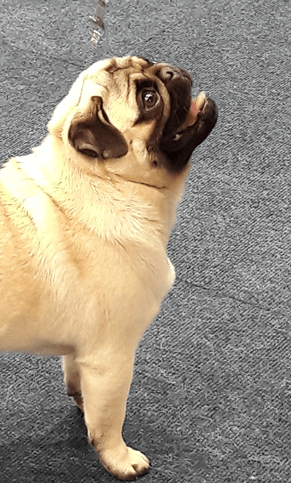
Pug.
Picture: QUEN archive
2.1 Picture 2
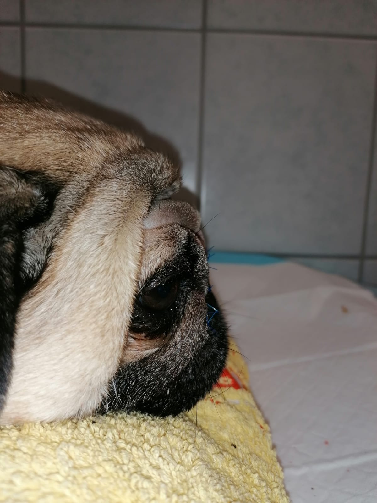
Pug after nasal fold surgery.
Picture: QUEN archive
More pictures can be found here (click on picture):
3. Problems/syndromes that may occur in the breed
The following breed-typical defects or common problems/health disorders and dispositions* are known in Pugs:
* (please also refer to the existing information sheets on individual defects such as brachycephaly and entropion in particular)
- Brachycephaly
- BOAS (brachycephalic obstructive airway syndrome)
- Tracheal collapse
- Spinal defects (spina bifida, vertebral body malformations, intervertebral disc disease (IVDD), myelopathies, shortened or deformed tail,
- Hip dysplasia
- Necrotising meningoencephalitis (pug encephalitis)
- Difficulties in labour (dystocia), high perinatal mortality
- Skin diseases (atopic dermatitis, skin fold dermatitis, demodicosis)
- Eye diseases (bacterial keratitis, cataract, corneal pigmentation, ulcerative keratitis (corneal ulceration), distichiasis, ectopic cilia, entropion, iris hypoplasia, keratoconjunctivitis sicca, macroblepharon, persistent pupillary membrane, sudden onset retinal degeneration – SARDS, traumatic proptosis (prolapsed eyeball))
- Ear diseases
- Misaligned teeth and other oral pathologies
- Mortality rate
4. Other problems that may occur frequently
In addition to the breed-typical defects listed under point 3, the veterinary literature contains information on the occurrence of the following problems, which will not be discussed further below, as no definitive conclusions can yet be drawn from the information on prevalence:
- Congenital portosystemic shunt
- Legg-Calvé-Perthes disease Patellar luxation
- Gear deviations
- Tumour diseases (mast cell tumours, squamous cell carcinoma of the cornea)
- Lung lobe torsion
- Aspiration pneumonia
- Overweight
- Urolithiasis (struvite stones)
- Kidney stones
- Pyometra
- Dental diseases (odontogenic cysts)
- Sick sinus syndrome (cardiac arrhythmia)
- Vaccine-associated adverse reactions
- Diabetes mellitus
- Seizures
- Pectus excavatum and pectus carinatum
5. Symptoms and pathological value of the typical defects mentioned above: Significance/impact on the physical/psychological well-being of the defect in the individual animal and classification in burden category*
*The individual breeding-related defects are assigned to different burden categories (BC) depending on their severity. The overall burden category is based on the most severe defect found in the individual animal. The BC system as a further development based on the Swiss model is still being developed and is only intended as a guide. For this reason, the BC values used here should be regarded as provisional. This is primarily due to the fact that the German Animal Welfare Act does not contain a justiciable basis for classification into burden categories. In contrast to Switzerland, the legal standards in Germany do not quantify pain, suffering or harm or assess their quality, but take them into account if they affect the animal more than insignificantly.
However, the burden categories can also be used to assess suitability for breeding and showing.
The burden that can be caused by defective breeding traits are divided into 4 categories (Art. 3 TSchZV, Switzerland). The most burdenful trait or symptom is decisive for the assignment of an animal to a burden category (Art. 4 TSchZV, Switzerland).
Category 0 (no burden): These animals may be used for breeding.
Category 1 (mild burden): Mild burden is present if a burdenful expression of characteristics and symptoms in pets and farm animals can be compensated for by appropriate care, husbandry or feeding, without interventions on the animal and without regular medical care measures.
Category 2 (medium burden): These animals may only be used for breeding if the breeding objective is for the offspring to be less burdened than the parents.
Category 3 (severe burden): These animals may not be used for breeding.
Brachycephaly
(see also https://qualzucht-datenbank.eu/merkblatt-hund-brachycephalie/)
Physical:
Brachycephaly describes in the broadest sense the shortening of the head, including the muzzle. The special anatomy of the upper airways in brachycephalic dogs leads to obstruction of the upper airways. Compared to other breeds, brachycephalic dogs have a shortened skull, which leads to a compression of the nasal passages and an altered pharyngeal anatomy, resulting in an altered anatomy of the upper and also partially the lower airways. This often restricts the function of the airways. When comparing the CT images of mesocephalic and brachycephalic dogs, it is noticeable that the facial skull of the pug itself is shortened in comparison with other brachycephalic breeds. In addition, it is shown how the intranasal malformations of brachycephalic facial skulls can result in aberrant, stenotic conchae nasales, which are often only slightly branched, and can obstruct both the rostral nasal passages and the choanae. This results in a reduction in the air supply.
According to a study, up to 90% of animals can be affected by malformed conchae nasales that obstruct the airways, especially rostrally. However, such malformed nasal conchae can also be present caudally until they partially obstruct the choanae. According to one study, up to 17% of Pugs suffer from an „extreme case“ in which the conchae nasales protrude into the pharynx and lead to obstruction of the airways. In the area dorsal to the palate, studies have shown that the airways of Pugs are relatively narrowed in their cross-section, even compared to other brachycephalic breeds. Most brachycephalic dogs have a combination of compressed anatomical structures that create increased negative pressure on inspiration, which leads to inflammation and stretching of the pharyngeal tissues, exacerbating obstruction, but can also have effects on the gastrointestinal tract, e.g. through reflux oesophagitis. Brachycephalic breeds have been shown to have frequent changes in the ear canal due to the shape of the skull. A malformation of the external opening of the auditory canal (porus acusticus externus) leads to severe narrowing of the proximal auditory canal. Independently of this, these animals also suffer more frequently from external and medial otitis. According to studies, the ratio of air to soft tissue within the naso-/oropharyngeal structures is up to 60% lower in brachycephalic dogs than in mesocephalic dogs. This is also due to the fact that brachycephalic dogs usually suffer from relative macroglossia due to their compact body conformation.
The volume does not differ between affected and healthy dogs, but the larger tongue in relation to the smaller skull also impairs breathing.
Very often, the nasolacrimal duct is malformed due to breeding-related changes to the facial skull and its course is very different. It is often considerably reduced in length and can have a steep slope. Of all brachycephalic dog breeds, the Pug has the greatest deviation from the physiological course. In many cases, the nasolacrimal ducts of the affected animals have an additional opening through which the fluid can also drain into the rear part of the nasal cavity.
Changes to the bony skull can also cause changes, diseases and an increased risk of injury to the eyes.
Brachycephaly is also associated with a malformation that often leads to changes in the dentition (brachygnathia superior).
Examinations show differences in the cardiac ECG between brachycephalic dogs and dogs without brachycephaly. In brachycephalic dogs, the diastolic pressure in the right heart appears to be increased, which impairs the function of the heart. The changes in cardiac morphology and function are usually determined by the anatomical changes, not by the appearance of symptoms associated with brachycephaly.
Psychological:
Many behaviours and areas of the animal’s life are negatively affected by brachycephaly and the resulting diseases. Respiratory distress may occur during feed intake (see explanations under BOAS). Affected animals usually require a longer recovery phase after short periods of activity. Playing with conspecifics may be restricted. Overall, it can therefore be assumed that the quality of life is significantly reduced. The altered anatomy of the skull also leads to changes in the musculature of the skull and the size of the eyes, so that the facial expressions and thus the communication of the animals with conspecifics is impaired. The flattened orbit associated with the skull alteration and the frequently existing macroblepharon often leads to protruding eyes, often combined with strabismus (squinting), which significantly impairs the animal’s visual acuity.
Burden category: 3
If the nose length is more than ⅓ of the total head length: Burden category 2
BOAS (brachycephalic obstructive airway syndrome)
Physical:
Brachycephalic airway syndrome is a combination of upper airway abnormalities in dogs that lead to upper airway obstruction. The shape of the skull has a major influence on the development of BOAS. The more pronounced the brachycephaly, the higher the risk of BOAS. The Pug is one of the breeds particularly affected and can have up to a 50-fold increased risk of BOAS compared to other breeds. The prevalence of BOAS in this breed in England was between 6.5 and 59.8%, depending on the study. In an Australian study, Pugs were presented with this problem at around two years of age and, at 26%, were the brachycephalic breed most frequently operated on due to BOAS symptoms. However, the affected animals are often not presented at the veterinary practice because the symptoms are not recognised by the pet owners. The prevalence may therefore be considerably higher. It is characterised by stenotic nostrils, an elongated soft palate, inverted laryngeal pockets and a hypoplastic trachea. Affected dogs can have any combination of these defects, usually three to four, leading to varying degrees of upper airway impairment. Nasal mucosal contact points that restrict airflow can exacerbate breathing problems. Due to the anatomical changes, brachycephalic dogs have a significantly increased respiratory resistance against which they have to breathe. The intraluminal pressure is increased during inspiration and is the cause of inflammation, inverted laryngeal pockets and possibly laryngeal and tracheal collapse. Clinical signs include inspiratory stertor (gasping breath), stridor, cough, exercise intolerance, choking, vomiting, syncope and dyspnoea. Extreme breathlessness may be associated with difficulty in feeding and uncoordinated swallowing, which can lead to aspiration pneumonia and aerophagia.
Vomiting, regurgitation and increased salivation are among the gastrointestinal symptoms that can occur in connection with BOAS. The reflux oesophagitis that often results can cause severe pain.
The lower airways are also affected in BOAS due to the increased respiratory resistance in the upper airways. Collapsed bronchi can be observed. After exercise and due to the sensitivity to heat (disruption of thermoregulation), the animals need significantly longer to recover. In an Australian study, the Pug was the breed with the highest incidence of laryngeal collapse, with a prevalence of 38%. Affected dogs can be recognised by snoring noises.
If the clinical signs are severe, surgical correction may be necessary with a corresponding risk of anaesthesia and possible postoperative complications.
Affected dogs can be impaired in all areas of life due to the consequences of brachycephalic conformation. They need a longer recovery period after physical activity. Sometimes the dogs can only sleep in certain positions in order to be able to breathe sufficiently and therefore suffer from various sleep problems.
The sleep disorders range from sleep apnoea and choking during sleep to adopting an upright sleeping position (raised head, attempts to sleep in a sitting position) or using objects in the mouth to facilitate breathing. The prevalence of sleep disorders caused by BOAS is characterised by a high number of unreported cases, as owners consider the signs to be normal and the affected animals are therefore often not recognised as such.
BOAS as a result of the brachycephalic head shape can lead to death.
Psychological:
Interactions with other dogs may be limited due to the breathing noises. The animals are severely restricted in many areas of life due to impaired physiological bodily functions and reduced fulfilment of basic needs, so that a reduced quality of life can be assumed. An alarmingly high proportion of these animals are unable to sleep and this also leads to an immense reduction in their quality of life.
Burden category: 3
Tracheal collapse
Physical:
Tracheal collapse is described as occurring in pugs as well as some miniature breeds. Tracheal collapse is a form of tracheal obstruction caused by cartilage laxity and flattening. In affected animals, the cartilage collapses in a dorsoventral direction, with the cervical trachea collapsing during inspiration and the thoracic trachea collapsing during expiration. Clinically, a chronic, paroxysmal cough is typically noticeable. However, the animals can also be asymptomatic. Compared to other dog breeds, the pug can have a relatively narrow tracheal ring at the thoracic inlet.
Psychological:
The dogs suffer from chronic, paroxysmal chesty cough with accompanying breathlessness and fear of suffocation. Tracheal collapse is a potentially life-threatening respiratory disease.
Burden category: 3
Spinal defects (tail deformities, spina bifida, vertebral body malformations, intervertebral disc disease (IVDD), myelopathies)
Physical:
Spinal deformities occur frequently in Pugs. They can be single or multiple and include hemivertebrae, spina bifida or transitional vertebrae. The diagnosis of vertebral malformations is often an incidental finding, especially in chondrodystrophic breeds. Studies show that over 80% of the non-symptomatic dogs of the Pug, Bulldog and French Bulldog breeds examined have at least one vertebral malformation, and around 60% of the animals also have multiple vertebral malformations. The vertebral malformations are not always accompanied by neurological symptoms. The prevalence of clinically relevant thoracic vertebral malformations was 4.7% in an English study of Pugs. Spina bifida occurred in another English study in the breed with a prevalence of 8.5 % and lumbosacral transitional vertebrae in 54.2 %. Breeds with a tail deformation may have a higher incidence of other spinal defects. However, the Pug does not carry the DVL 2 gene responsible for the „screw-tail“ and the pathoetiology of vertebral malformations may therefore differ from that of the French Bulldog, Boston Terrier and Bulldog.
(See QUEN data sheet no. 5 Dog Tail (Fact sheet Dog Tail – QUEN Qualzucht-Database) and Fact Sheet No. 24 Dog Robinow-Like Syndrome (Fact Sheet Dog Robinow-Like Syndrome – QUEN Qualzucht-Database (qualzucht-datenbank.eu)).
Hemivertebrae and Intervertebral Disc Disease (IVDD)/ Intervertebral Disc Herniation (IVDH)
Hemivertebrae occur more frequently in chondrodystrophic breeds such as the pug. The breed-specific prevalence is between 1.7 % and 17.6 %, depending on the study. Hemivertebrae can occur unilaterally or bilaterally. In Pugs, ventral hypoplasia of the vertebral bodies is predominant even in clinically normal animals and is associated with larger Cobb angles and a higher probability of kyphosis. A correlation was also found between spinal kyphosis with a Cobb angle of > 35° and neurological deficits. The Cobb angle is measured using X-ray images and indicates the degree of curvature of the lateral or sagittal spinal curvature.
Affected animals are usually less than one year old and can be clinically inconspicuous. In the Pug, however, the occurrence of hemivertebrae may be associated with the development of clinical signs to a greater extent than in the French Bulldog and Bulldog. This is also attributed to vertebral instability, among other things. Hemivertebrae can lead to paresis, ataxia, paraplegia with intact nociception, kyphosis and, more rarely, spinal hyperaesthesia and urinary and faecal incontinence. There is a risk of spinal cord compression, which can lead to corresponding neurological symptoms. Breed-specific vertebral body malformations in Pugs also include transitional vertebrae, for which the risk was approximately 22 times higher than in French Bulldogs in an Australian study. In an English review study, in which CT scans of different breeds were analysed, 46% of Pugs had cervical ribs, which were frequently associated with thoracolumbar transitional vertebrae. According to studies, Pugs suffer more frequently from kyphosis than other extremely brachycephalic breeds. Due to their influence on the biomechanics of the spine, kyphoses are usually associated with neurological symptoms or IVDD. The severity of kyphosis is thought to be related to the likelihood of neurological symptoms occurring in dogs affected by hemivertebrae.
There is evidence that anomalies of the caudal vertebral articular process are frequently associated with herniated intervertebral discs (IVDH) in the thoracolumbar region. In pugs, the articular processes were hypoplastic or aplastic, i.e. underdeveloped or not present at all, in over 90% of the cases examined. The resulting neurological deficits can be acute or chronic.
In a Japanese study, the prevalence of IVDH was 2.4% of dogs in the thoracolumbar region and 0.8% in the cervical region.
Degenerative myelopathies (DM)
According to the Orthopedic Foundation for Animals (OFA), degenerative myelopathy in Pugs is present in 11.8% of the animals tested and of these animals, approx. 33% were carriers of the gene. In an English referral clinic, the prevalence was 64.9% of the cases analysed. Animals with thoracolumbar myelopathy often show a chronic, progressive clinical course with ataxia and paraparesis, often accompanied by incontinence. Gait disturbances caused by myelopathies are a common problem in Pugs. Affected animals are on average seven years old when they first show neurological symptoms.
Insurance data from a large pet insurance company, Agria Pet Insurance Sweden, has shown that in Sweden the mortality rate for Pugs with neurological symptoms such as ataxia and paresis is approximately 5 times higher than the average mortality rate for all breeds, and in Sweden affected animals usually have to be euthanised within a year of the onset of ataxia.
A mutation in the SOD1 gene has been identified in Pugs that is associated with degenerative myelopathy. Approximately 30% of Pugs tested in a study on the frequency of the disease in different breeds were carriers of the mutation and 17.5% of the animals had a high risk of developing degenerative myelopathy. A more recent study identified 34 genes, 14 of which have functions that could be linked to the pathogenesis of DM.
Psychological:
The clinic described above due to vertebral deformities severely restricts the animal’s well-being due to pain, and slipped discs are often accompanied not only by signs of paralysis but also by severe pain, which significantly impairs the animal’s well-being. A deformed tail or a tail that is missing except for a few limbs can hardly or no longer be used as a means of expressing the natural and species-appropriate behavioural repertoire. Restrictions to the behavioural repertoire are recognised as suffering, also because in this case it massively restricts communication with conspecifics.
Burden category: 3
Hip dysplasia
Physical:
Hip dysplasia is also listed among the predispositions in Pugs, with a prevalence of up to 69.7 % being assumed in some cases. The OFA test statistics show a prevalence of 42.9 % mild, 23.2 % moderate and 14.3 % severe hip joint dysplasia in Pugs. The clinical picture can vary. The function of the hind limb can be impaired depending on the changes and progression. Signs of pain, lameness and restricted movement may be part of the clinical picture. The dogs either present with hip instability at a young age or with advanced osteoarthritis in adulthood.
Psychological:
The pain and lameness or restricted movement can restrict the animals in their active, species-typical behaviour and impair their well-being.
Burden category: 2-3
Necrotising meningoencephalitis (NME) (pug dog encephalitis (PDE))
Physical:
The prevalence of PDE in pugs is approx. 1-2%. The underlying aetiology of idiopathic necrotising meningoencephalitis is not yet clear. A hereditary autoimmune disease is being discussed and high-risk alleles on certain gene loci have been identified. In Europe, the frequency of high-risk alleles in Pugs is 25.7%. The prevalence of the homozygous high-risk gene mutation is 7.4%.
The majority of Pugs with necrotising meningoencephalitis had multifocal or diffuse, asymmetrical lesions in the forebrain (prosencephalon), although at least one lesion was located in the cerebrum (telencephalon). Two thirds of dogs also have a diencephalic lesion. Sometimes lesions can also be detected in the cerebellum and brain stem. The dogs can become conspicuous due to (generalised) convulsions. Anorexia, circling, tremor, paresis and behavioural changes may also occur. Inflammatory and necrotic changes are also associated with the severity of the clinical picture. Histopathologically, dilatation of the lateral ventricles and multifocal or laminar necrosis of the cerebral cortex can be observed. Colour abnormalities, malacia and cavitations are also part of the pathology. At the time of diagnosis, the dogs are usually around two years old. The disease is often fatal for the animals.
Psychological:
The disease can lead to pain and suffering.
Burden category: 3
Birth difficulties (dystocia), high perinatal mortality
Physical:
Brachycephalic breeds are particularly at risk of developing dystocia. Due to the brachycephalic skull shape, the heads of the foetuses are often too large in relation to the pelvic dimensions and the Pug is also one of the breeds that most frequently have a particularly narrow pelvic inlet. In a study from Great Britain, the Pug was the third most commonly affected breed with dystocia, accounting for 6.1 % of all breeds. The prevalence within the breed there was 14.5 % between 2012 and 2014. In Sweden, the Pug is in fifth place for the highest incidence of dystocia. Birth difficulties are often associated with a caesarean section, i.e. a surgical procedure. Labour difficulties can become an emergency, which is risky for the bitch and the puppies. In a Norwegian study, the risk of perinatal mortality in Pugs was 17%. Perinatal mortalities can include stillbirths and mortalities during the first seven days of life.
Psychological:
Caesarean section can influence maternal behaviour. Vaginocervical stimulation appears to play an important role in maternal behaviour, as it has been observed that cesarean section bitches may have problems developing adequate interactions with their puppies without the induction of natural birth.
Burden category: 3
Skin diseases
Physical:
Brachycephalic breeds are often affected by skin diseases, with the Pug being one of the particularly predisposed breeds. The prevalence of skin diseases in Pugs in England between 2009 and 2015 was 15.6 %. The risk of developing skin fold dermatitis (intertrigo) can be approx. 10 to 16 times higher compared to other breeds. Reduced air circulation and increased temperature, moisture and debris in the skin folds, together with intermittent friction and resulting microtrauma, lead to overgrowth of commensals such as microorganisms, bacteria and fungi and the production of toxins, followed by inflammation, maceration and infection. The affected areas may show erythema, hypotrichosis, alopecia, erosion/ulceration, crusting, lichenification (extensive leathery changes to the skin), pigmentary changes, accumulation of debris and bad odour. Skin folds at the base of the morphologically altered tail lead to secondary infections of the skin. If no surgical intervention is performed, lifelong treatment with topical substances may be necessary. In severe cases of demodicosis, bacterial infections can lead to septicaemia and non-specific symptoms such as fever, anorexia, lethargy and peripheral lymphadenopathy.
Some skin diseases, such as skin fold dermatitis, are directly linked to the abnormal conformation of brachycephalic dogs. Other skin diseases, such as atopic dermatitis and viral pigmented plaques caused by papillomaviruses, have a genetic basis or a general predisposition. Anatomical changes associated with brachycephaly, which lead to wrinkling of the skin and narrowing of the ear canal, favour the development of pyoderma, Malassezia dermatitis and otitis externa/interna (see ear diseases).
Atopic dermatitis is a hypersensitivity to allergens in the environment. Atopic dermatitis also increases the risk of immunoglobulin E-mediated food allergies. The symptoms can initially be seasonal and increasingly occur all year round. Ventral areas, armpits, groin and interdigital areas as well as the area around the muzzle, eyes and ears are frequently affected. Atopy is usually characterised by itching; infections can follow.
Demodicosis is a parasitic disease to which the Pug is predisposed. Demodex mites can be found in small numbers on almost all dogs. In young dogs, an insufficiently mature immune system (T-lymphocyte defect) may contribute to the development of demodicosis. Multifocal hypotrichosis, including alopecia, erythema, crusts, scales, follicle formation, papules, pustules, nodules, hyperpigmentation, lichenification and comedones on the head, trunk, limbs and paws occur in varying degrees. Secondary infections, especially with bacteria, are common and can lead to itching.
Psychological:
Many skin conditions can become chronic, causing pain and itching, which can lead to behavioural problems and affect quality of life. Skin fold dermatitis may (initially) go unnoticed by the owner, as the changes are located between the skin folds. Depending on the severity of the disease, the animals‘ well-being may be significantly impaired by the constant itching alone.
Burden category: 2-3
Eye diseases
Physical:
Brachycephalic breeds have anatomical skull abnormalities that are responsible for clinical eye symptoms. The brachycephalic ocular syndrome (BOS) summarises a number of eye abnormalities that occur in combination with brachycephaly. The frequently occurring ocular anomalies include corneal ulcers (corneal ulcers/ulceration or ulcerative keratitis), corneal pigmentation, corneal fibrosis and entropion. A bilaterally occurring enlarged palpebral fissure (macroblepharon) can also frequently be observed. Studies by the University of Vienna have shown that macroblepharon is often associated with strabismus. In an American study, the Pug was one of the dog breeds most frequently affected by eye diseases, accounting for 20.8% of cases. Within the breed, 16.25 % of dogs can be affected by eye diseases.
Pugs are particularly prone to corneal lesions. The Pug is one of the most common breeds to develop corneal ulcers, with a prevalence of approx. 5%. Depending on the data available, the risk of corneal ulcers in Pugs is approx. 10 to 13 times higher than in other breeds. Reduced protective mechanisms, for example through protrusion of the eyes, are described as a favouring factor. The disease can be associated with pain for the affected dogs. A five times higher risk of corneal ulcers has been described for dogs with nasal folds, and for brachycephalic dogs with a craniofacial ratio < 0.5 even twenty times higher than for non-brachycephalic breeds. The craniofacial ratio describes the muzzle length divided by the skull length and quantifies the degree of brachycephaly.
The number of affected dogs with keratitis pigmentosa varies greatly between the studies. It ranges from 22.9 % to 53 % to 71.8 % and 82.4 %. Corneal pigmentation develops in connection with chronic inflammation, which can spread to the visual axis. In a study from Great Britain, 91.9 % of the participating Pugs were diagnosed with keratitis pigmentosa. Of the affected dogs, 91.2 % were affected on both sides. Detection was significantly associated with older age and the presence of medial entropion of the lower eyelid. There was also a correlation between the severity of keratitis pigmentosa and the degree of medial entropion of the lower eyelid. The aetiology is not yet fully understood.
Other eye changes occur in connection with BOS, but these are not exclusively BOS-specific. Known or suspected hereditary eye problems include trichiasis, caruncle trichiasis, distichiasis, ectopic cilia, entropion, corneal dystrophies, prolapse of the nictitating gland, keratoconjunctivitis sicca, hereditary cataract and progressive retinal atrophy as well as reduced corneal sensitivity. The exophthalmos prevents effective blinking, which leads, among other things, to drying of the eyes with a loss of corneal sensitivity. The flattening of the bony orbit and the enlarged palpebral fissure can lead to prolapse of the globe (traumatic proptosis), which can result in loss of the eyeball if no immediate intervention is taken.
Diseases such as keratoconjunctivitis sicca occurred in 9.1 % of dogs in an American evaluation. The breed can also be affected by a primary (2.28 %) or immature cataract (4.1 %). Corneal perforation occurred in 4.3% of the dogs. Up to 72.1% of Pugs can be affected by iris hypoplasia. In a prospective study, 83.8 % or 85.3 % of Pugs had a persistent pupillary membrane on one or both sides.
In a retrospective study, the Pug was one of the most common breeds for bacterial keratitis, accounting for 8% of cases. It is assumed that the protective mechanisms of the eye are impaired in brachycephalic breeds and that exophthalmos, skin folds and entropion favour bacterial keratitis. S. intermedius, β-hemolytic Streptococcus spp. and P. aeruginosa were most frequently isolated.
In a comparison of 60 dog breeds, the Pug ranked third in the frequency of sudden acquired retinal degeneration (SARD). The Pug was also over-represented in other studies for this disease. The dogs were presented due to (mostly sudden) blindness.
Overall, excessive breeding selection leads to extreme skull shapes, which in turn lead to facial changes and jeopardise the dogs‘ eyesight. Surgical interventions are often necessary in dogs with BOS. Excessive nasal folds must be removed to prevent trichiasis, for example. Diseases such as pigment keratitis can have a negative effect on vision. If the inflammation in pigment keratitis spreads to the visual axis, this can lead to considerable visual impairment and, in severe cases, blindness. The frequently occurring strabismus also impairs vision in a pronounced form.
Psychological:
The eye diseases can cause pain in the animals and, depending on the severity, restrict their ability to see. Severely visually impaired or blind dogs are barely able to use body signals such as posture, tail position or eye, head and mouth signals or to recognise them in other dogs. This can lead to changes in behaviour, anxiety and insecurity.
Burden category: 2-3
Ear diseases
Physical:
The proximal external auditory canal in brachycephalic breeds such as the pug is characterised by a clear stenotic malformation. Narrowing and reduction of the lumen by up to 50 % can be observed. The prevalence of otitis externa is significantly higher in brachycephalic breeds such as the Pug, but varies between 7.53% and 32% depending on the study. Various factors are associated with otitis externa. In addition to the narrowing of the ear canal, the accumulation of soft tissue and fluid in the lumen of the ear canal can be a favouring factor. Although malformation of the external auditory canal in brachycephalic dogs can cause severe stenosis of the proximal auditory canal, a recent study has not confirmed a link with otitis externa.
Brachycephalic dogs tend to accumulate fluid in the middle ear (tympanic cavity effusion). This is attributed to subclinical otitis media in connection with otitis externa or to drainage disorders due to an abnormal morphology of the tympanic cavity.
A tympanic cavity effusion is often diagnosed in Pugs as an incidental finding during CT examinations. Depending on the study, the prevalence is between approx. 16% and 25%. Cytological and bacteriological examinations of the secretions showed high concentrations of inflammatory cells. However, an effusion without signs of otitis media may also be present. Bacterial infections are only present in some cases and are usually limited to the detection of Staph. pseudointermedius .
The stenosis and fluid build-up can cause hearing loss, which can lead to deafness in the affected ear.
Psychological:
The narrowing of the ear canal makes the treatment of otitis media and externa more difficult, as it is difficult to examine and visualise the structures, such as the eardrum. Relapses after treatment are possible, so that the animals then have to undergo long therapies and repeated examinations (often only possible under anaesthetic). Otitis can be very painful and, if it becomes chronic, can restrict the animal’s quality of life.
Burden category: 2
Misaligned teeth and other oral pathologies
There are not too many studies on oral pathologies, malpositions of the jaws and teeth and their frequency. The occurrence of these and other defects in brachycephalic breeds now seems to be accepted as normal.
Frazer A. Hale comments on this with an observation that applies to many hitherto little-studied breeding-related defects that are either a direct consequence of the deformed facial anatomy or the result of other genetic defects associated with the brachycephalic genotype: „The absence of evidence is not the evidence of absence“.
Due to the altered/compressed anatomy of the facial skull, deviations from the normal state and associated peridontal diseases are regularly found in the dentition of brachycephalic animals, albeit in varying degrees of severity and stage of progression.
Physical:
Dental crowding and rotation
In the severely shortened upper jaw of brachycephalic dogs, there is not enough space to accommodate the physiological number of 20 teeth (ten on each side) in the necessary distance and arrangement/position. This leads to various health problems, which are essentially caused by the fact that the gingiva – the physical barrier between the bacteria in the oral cavity and the jaw bones that hold the tooth roots – that normally surrounds each tooth is not intact.
In pugs, for example, the premolars are typically twisted by 90%, which often leads to tooth loss due to inflammation with subsequent loss of bone substance around the tooth roots.
However, the lower jaw is also too short in relation to the head, so that malocclusions can also occur there with the same consequences for the oral health of the oral cavity and thus also for general health.
Malocclusion with abnormal tooth contact
All brachycephalic animals have a malocclusion of the third degree (required by the breed standard) because the upper and lower jaw lengths diverge in relation to each other.
The only place in the canine mouth where contact between teeth in the upper jaw and other teeth in the lower jaw should physiologically occur is where the two upper molars make contact with the distal third of the first mandibular molar and the second and third mandibular molars. Any other contact of teeth in the canine mouth is a deviation and undesirable. In addition, the gingiva should not overlap/overgrow the teeth. It is also frequently observed that the incisors of the upper jaw meet the lingual side of the incisors of the lower jaw or their gingiva. The animals bite themselves every time they close their mouths. It is not uncommon for healthy teeth to have to be proactively extracted to compensate for the breeding-related malformation.
Non-erupting teeth and odontogenic cysts
Odontogenic cysts are fluid-filled structures that can form over the crown of unerupted teeth.
These odontogenic cysts can occur in all dogs, but are observed much more frequently in brachycephalic animals. The absence of teeth in the permanent dentition of young dogs is an indication for an intraoral X-ray examination of the dentition.
Ulcerative palatitis induced by foreign bodies
In brachycephalic dogs, the palatal rugae on the mucosa are strongly folded, comparable to a folded harmonium. To prevent the accumulation of hair, foreign bodies, debris and bacteria, the oral cavity must be checked daily and cleaned if necessary.
Psychological:
Overall, it should be noted that dogs, like cats, are known not to show pain in this area and do not refuse to eat despite sometimes considerable defects and illnesses, because the feeling of hunger prevails as a self-preservation instinct.
This means that pain and suffering can be present without being recognised by the dog owner.
Burden category: together with brachycephaly: 3
Mortality
Data from a large Swedish insurance company shows that the Pug has a 4-fold increased risk of mortality due to neurological diseases, followed by a 3.5-fold increased risk due to eye diseases, compared to the average mortality rate of all breeds.
In the case of the Pug, the breed-related (standard-related) initial values already result in the overall category 3.
6. Heredity, genetics, known gene tests, if applicable, average coefficient of inbreeding (COI), if applicable, Generic Illness Severity Index
Brachycephaly (shortening of the facial skull and associated problems with thermoregulation, ear disorders, etc.)
Genetics and inheritance are not fully understood. Due to the genetic complexity, it is assumed that various chromosomes have an influence. The genetic and developmental basis of brachycephaly is complex and involves multiple gene regulatory networks, reciprocal signalling interactions and hierarchical levels of control. Several studies have found links to the CFA1 chromosome. The involvement of SMOC2, BMP3 and DVL2 genes is also being discussed.
A genetic test is not available.
PDE
Due to the high degree of heritability, genetic causes are being discussed as well as (auto)-immunological factors and the influence of environmental factors. Two important disease-associated gene loci have been identified, similar to the histocompatibility complex (MHC) loci previously identified in MS. The haploid genotype susceptible to the disease significantly increases the risk of developing PDE in homozygous individuals. An autosomal recessive inheritance with variable penetrance is suspected.
A genetic test is available.
Degenerative myelopathy (DM)
Degenerative myelopathy, which is caused by a mutation in the SOD1 gene, is an inherited neurological disorder in dogs. This mutation occurs in many dog breeds, including the Pug. The inheritance of DM is autosomal recessive with age-dependent incomplete penetrance.
A genetic test is available.
Other genetic tests available for the breed (as of October 2024):
- Chondrodysplasia and dystrophy (IVDD risk)
- Hyperuricosuria
- Malignant hyperthermia
- May-Hegglin anomaly
- Primary lens luxation
- Progressive retinal atrophy (prcd-PRA)
- Pyruvate kinase deficiency (PK)
Average inbreeding coefficient of the breed:
The coefficient of inbreeding (COI) for the Pug was over 40% on average when analysing the SNPs . This value is higher than for a mating of half-siblings, which leads to an inbreeding coefficient of 12.5 % in the puppies, and even higher than a mating of two full siblings, in which the inbreeding coefficient of the puppies would be 25 %.
7. Diagnosis – necessary examinations before breeding or exhibitions
Caution: Invasive examinations that are stressful for the animal should only be carried out in justified cases of suspicion in breeding animals and not if visible defects already lead to a ban on breeding and showing.
Brachycephaly
In vivo measurements can be used to determine the extent of brachycephaly. Brachycephalic dogs are characterised by a skull width of at least 80% of the skull length. A skull width of more than 80 % of the length is considered extremely short-snouted. Brachycephalic syndrome can be diagnosed using imaging techniques. Craniometric measurements can be taken with X-rays. The upper airways can be visualised using CT and endoscopy. A practical method of measuring the skull was developed in the Netherlands using the traffic light system. Breeding is prohibited there in the case of relative nasal shortening with a craniofacial ratio of less than 0.3, i.e. the nasal length must be at least ⅓ of the total head length.
BOAS (brachycephalic obstructive airway syndrome)
During the anamnesis, attention should be paid to the corresponding clinical symptoms and previous history. To assess a possible BOAS, the dogs can be examined clinically before and after exercise. The nostrils and any forced breathing can be examined. By dividing the nasal length (cm) by the skull length (cm), the craniofacial ratio (CFR) or relative nasal shortening can be calculated. This can be used to determine the severity of BOAS. For a more detailed examination of the anatomical structures, imaging procedures such as an X-ray, CT or endoscopy can be performed under anaesthesia. Further examinations such as plethysmography are possible, but further findings are essential to determine whether an animal may be bred or exhibited. The so-called Cambridge Respiratory Function Grading Scheme (RFGS) was introduced by breeding associations as a standard for breeding recommendations with regard to BOAS-related health problems in Pugs, Bulldogs and French Bulldogs. The findings and results obtained from this do not take into account any other breeding-related defects and are not suitable on their own for determining suitability for breeding or showing. According to a breeding association, „functional tests such as the Cambridge test have shown better significance than, for example, the craniofacial ratio (ratio of nose length to skull length) used in the Netherlands“.
However, such a test is a snapshot in time of usually young animals and ignores the fact that the probability of the occurrence of symptoms from the BOAS increases with increasing age and also with obesity, which is common in Pugs, and that a number of other defects in the teeth, ears, eyes, nasolacrimal canal and thermoregulation are regularly associated with the main symptom brachycephaly.
Tracheal collapse
A characteristic chronic cough should be noted in the medical history. Radiological imaging may reveal a narrowed tracheal lumen. The cough occurs mainly during excitement, activity or food intake. The symptoms are described as chronic. Auscultation can be used to make an initial assessment of the possible localisation. The trachea should be palpated carefully. Tracheal collapse is a dynamic process, so attention should be paid to whether the animal is in inspiration or expiration when analysing the X-ray. Fluoroscopy or bronchoscopy can be used for further assessment.
Birth difficulties (dystocia)
During the anamnesis with clinical examination and questioning about the pregnancy and the birth process, attention should be paid to certain findings, such as pelvic fractures, duration of pregnancy, body temperature and time between the birth of the puppies. A digital vaginal examination should be included. X-rays provide information about the number and position of the puppies.
Skin diseases
Depending on the suspected diagnosis, the anamnesis and clinical examination are followed by appropriate further examinations.
The different stages of Demodex mites can be identified, for example, by a deep skin scraping, a trichiogram or an acetate tape impression technique.
Spinal defects (spina bifida, vertebral body malformations, intervertebral disc disease – IVDD)
Multidimensional imaging procedures are suitable for diagnostics in order to be able to assess the spine. Depending on the health condition of the dog, also with regard to possible upper respiratory problems, anaesthesia may be safer than sedation during imaging. A gene test to rule out the risk of IVVD can be performed.
Eye diseases
A complete ophthalmological examination should be carried out before breeding or showing and the various parts of the eye should be examined.
Hip dysplasia
The dog is assessed at rest, at a walk and at a trot. In addition to a clinical examination, an orthopaedic examination is also carried out to assess the hip joint and radiological imaging enables a detailed assessment.
Necrotising meningoencephalitis
The diagnosis is based on an anamnesis with clinical history, clinic and histological examinations. Imaging using MRI or CT can visualise the occurrence and distribution of necrosis.
Ear diseases
Diagnostic measures include video otoscopy, myringotomy and MRI or CT. Otoscopic examinations of the auditory canals and the eardrum can be difficult due to constrictions. A more precise assessment of the auditory canals or the tympanic bulla is possible using CT.
8. Necessary or possible orders from an animal welfare perspective
Decisions on breeding or show bans should be made in connection with the burden category (BC). Depending on the severity and findings, the decisive factor for a breeding ban may be the most severe finding, i.e. the one that affects the animal the most, and its classification in one of the burden categories, or also the correlation assessment if there are many individual breeding-related defects. The individual genetic inbreeding coefficient of an animal should also be taken into account.
a) Orders that appear necessary
Breeding ban according to §11b TierSchG for animals with hereditary/breeding-related defects of stress categories 2 and 3, in particular with
- Brachycephaly with nasal length less than ⅓ of the total head length, because this is usually associated with further defects, in this respect brachycephaly is to be understood here as a leading symptom, which must always induce further examinations of the animal.
- Changes in the skeletal system (head, spine, hips, pelvis)
- Brachycephalic obstructive airway syndrome (BOAS)
- Extremely shortened upper jaw, malpositioned teeth or malocclusion, absence of several molars
- hereditary eye diseases or strabismus
- Obstructions of the nasolacrimal canal
- Pug Dog Encephalitis
Exhibition ban according to § 10 TierSchHuV
- Brachycephaly with a nasal length of less than ⅓ of the total head length, because this is usually associated with other defects in other areas, such as teeth, jaw, eyes, ears, palate, respiration, thermoregulation.
b) Possible orders
Order for permanent infertility (sterilisation/castration) in accordance with Section 11b (2) TierSchG
Please note:
Measures taken by the competent authority must be recognizably suitable for averting future harm to the animal concerned and/or its offspring. With regard to the type and depth of processing of orders and breeding bans, decisions are always made on a case-by-case basis at the discretion of the competent authority, taking into account the circumstances found on site.
9. General assessment of animal welfare law
a) Germany
From a legal point of view, dogs with the defects/syndromes described above are categorised as torture breeding in Germany in accordance with § 11b TierSchG.
Justification:
According to § 11b TierSchG, it is prohibited to breed vertebrates if breeding knowledge indicates that, as a result of breeding, the offspring or progeny will, among other things
- body parts or organs are congenitally missing or unsuitable or deformed for use in accordance with the species and this results in pain, suffering or damage (§ 11b Para. 1 No. 1 TierSchG) or
- the keeping is only possible with pain or avoidable suffering or leads to damage (§ 11b Para. 1 No. 2 c TierSchG).
The International Association for the Study of Pain (IASP) defines pain as
„an unpleasant sensory and emotional experience associated with or resembling actual or potential tissue damage (https://www.iasp-pain.org/wp-content/uploads/2022/04/revised-definition-flysheet_R2-1-1-1.pdf)
Pain is defined in animals as an unpleasant sensory perception caused by actual or potential injury, which triggers motor or vegetative reactions, results in learned avoidance behaviour and can potentially change specific behaviours (Hirt/Maisack/Moritz/Felde, TierSchG, Kommentar 4th ed. 2023 § 1 para. 12 mwN; basically also Lorz/Metzger TierSchG 7th ed. § 1 para. 20).
Suffering is any impairment of well-being not already covered by the concept of pain that goes beyond simple discomfort and lasts for a not insignificant period of time (Hirt/Maisack/Moritz/Felde Tierschutzgesetz Kommentar 4th ed. 2023 § 1 para. 19 mwN; Lorz/Metzger, TierSchG Komm. 7th ed. 2019 § 1 para. 33 mwN). Suffering can also be physically and mentally debilitating; fear in particular is categorised as suffering in the commentary and case law (Hirt/Maisack/Moritz/Felde Section 1 TierSchG para. 24 mwN; Lorz/Metzger Section 1 TierSchG para. 37).
Damage occurs when the physical or mental condition of an animal is temporarily or permanently altered for the worse (Hirt/Maisack/Moriz/Felde TierSchG Komm. 4th ed. 2023 § 1 para. 27 mwN; Lorz/Metzger TierSchG Komm. 7th ed. 2019 § 1 para. 52 mwN), whereby completely minor impairments based on a physical or psychological basis are not taken into account. „The target condition of the animal is assessed on animals of the same species. The absence of body parts is regularly assessed as damage in the commentary literature“ (VG Hamburg decision of 4 April 2018, 11 E 1067/18 para. 47, also Lorz/Metzger TierSchG Komm. § 1 para. 52).
The breeding of Pugs constitutes torture breeding due to the single or multiple damages, pain and suffering described in detail in section 5:
- Brachycephaly and associated pain and damage
- Damage to the spine and associated pain
- Damage to the hip joints (hip dysplasia) and associated pain,
- Eye diseases and associated pain, suffering and damage
- Skin diseases and associated pain, damage and suffering
- Ear diseases and associated pain, damage and suffering
- Inability to bear offspring naturally
- Impairment of thermoregulation and associated suffering
- Anxiety caused by shortness of breath
- Suffering due to limited ability to communicate facial expressions
- Fulfilment of the concept of suffering due to physical changes brought about by breeding measures, which lead to a not insignificant impairment of the species-specific behavioural processes and possibly non-resilience of the animals. A species-appropriate life is thus not insignificantly impaired and well-being is severely restricted.
It should be noted that a breeding ban applies not only when breeding is carried out with animals that themselves have traits relevant to cruelty (trait carriers), but also when it is known or must be known that an animal used for breeding can pass on traits that can lead to one of the detrimental changes in the offspring (carriers, in particular animals that have already produced damaged offspring; Lorz/Metzger, Kommentar zum TierSchG § 11b Rn. 6 with further evidence).
– An important indication of a hereditary defect is that a disease or behavioural abnormality occurs more frequently in related animals than in the overall population of the dog species. The fact that the breed or population has proven to be viable over a longer period of time does not speak against damage (see Lorz/Metzger commentary on the TierSchG § 11b para. 9).
– The prohibition applies regardless of the subjective facts, i.e. regardless of whether the breeder himself recognised or should have recognised the possibility of harmful consequences. Due to this objective standard of care, the breeder cannot invoke a lack of subjective knowledge or experience if the respective knowledge and experience can be expected from a careful breeder of the respective animal species (see Hirt/Maisack/Moritz, Tierschutzgesetz, Kommentar, 4th ed. 2023, Section 11b TierSchG para. 6).
– Inheritance-related changes in the descendants are also foreseeable if it is uncertain whether they will only occur in later generations after a generational leap (cf. Goetschel in Kluge § 11b para. 14).
b) Austria
Dogs with the defects/syndromes described above are categorised as torture breeding in Austria in accordance with § 5 TSchG
In particular*, it is a violation of Section 5 of the Austrian Animal Welfare Act to „carry out breeding which is foreseeable to be associated with pain, suffering, harm or fear for the animal or its offspring (torture breeding), so that as a result, in connection with genetic abnormalities, in particular one or more of the following clinical symptoms occur in the offspring not only temporarily with significant effects on their health or significantly impair physiological life courses or cause an increased risk of injury“.
*The word „in particular“ means that the list is not exhaustive but exemplary.
Breeding with dogs that suffer from the following defects and the associated problems or are genetically predisposed to them is to be qualified as torture breeding, as the following symptoms listed in § 5 are realised: Shortening of the facial skull (e.g. respiratory distress, malformation of the dentition), alteration of the external auditory canal and middle ear (e.g. deafness, neurological symptoms), ectropion and/or entropion (inflammation of the conjunctiva and/or cornea, blindness), vertebral body malformations and shortened tail (movement anomalies), skin fold dermatitis (inflammation of the skin), difficult births/caesarean sections.
The breeding of Pugs already qualifies as torture breeding due to the fact that „it must be assumed with a high degree of probability that natural births are not possible“ (Österreich. TSchG, 2022).
c) Switzerland
Anyone wishing to breed with an animal that exhibits a trait or symptom that may lead to moderate or severe burden in connection with the breeding objective must first have a burden assessment carried out. Only hereditary burdens are taken into account in the burden assessment (see Art. 5 of the FSVO Ordinance on Animal Welfare in Breeding (TSchZV)). Dogs with defects that can be assigned to burden category 3 are subject to a breeding ban in accordance with Art. 9 TSchZV. It is also prohibited to breed with animals if the breeding objective results in category 3 defects in the offspring. Breeding with animals in category 2 is permitted if the breeding objective is for the offspring to be less affected than the parents (Art. 6 TSchZV). Annex 2 of the TSchZV lists characteristics and symptoms that can lead to moderate or severe burden in connection with the breeding objective. Degenerative joint changes, skull deformities with disabling effects on the ability to breathe, the position of the eyes and the birth process, excessive wrinkling, slipped discs, eye dysfunctions, persistent entropion and cataracts are explicitly mentioned. In addition, individual breeding forms are expressly prohibited in accordance with Art. 10 TSchVZ. In other cases, however, a breeding ban is only imposed on a case-by-case basis. Animals that have been bred on the basis of unauthorised breeding objectives may not be exhibited (Art. 30a para. 4 let. b TSchV).
d) Netherlands
It is forbidden in the Netherlands to breed dogs with a muzzle shorter than one third of the length of the skull, in accordance with Article 3.4 „Breeding with domestic animals“ of the Animal Keeper Decree and Article 2 „Breeding with brachycephalic dogs“ of the Decree.
Detailed legal assessments and/or expert opinions, if already available, can be made available to veterinary offices for official use on request.
10. Relevant jurisdiction
- Germany: Not known
- Austria: Provincial Administrative Court of Styria, exhibition ban, 7 March 2023, 41-3852, Provincial Administrative Court of Linz, dismissal of complaint against exhibition ban, 2 November 2023, LVwG-000544/100/SB
11. Order example available?
Not yet for the Pug, but for several other brachycephalic breeds (see French Bulldog fact sheet).
Examples of orders are only made available to veterinary offices for official use on request.

12. Bibliography/ References/ Links
Only a selection of sources on the defects described above and, where applicable, general literature on breeding-related defects in dogs is given here. More comprehensive literature lists on the scientific background will be sent exclusively to veterinary offices on request.
Note: The description of health problems associated with the trait, for which there is not yet sufficient scientific evidence, is based on the experience of experts from veterinary practice and/or university institutions as well as publicly accessible databases or publications from animal insurance companies and therefore originates from different evidence classes.
As breeding and showing are international nowadays, the information does not usually only refer to the prevalence of defects or traits in individual associations, clubs or countries.
AGRIA Pet Insurance, Sweden. (n.d.). Pug Agria Breed Profiles Life 2016-2021.
AGRIA Pet Insurance, Sweden. (n.d.). Pug Agria Breed Profiles Veterinary Care 2016-2021.
Bach, J.-P. (2023). Successful introduction of the „Cambridge Test“ in Germany [Verband für das deutsche Hundewesen e.V. (VDH)]. https://tierschutz.vdh.de/fileadmin/VDH/media/tierschutz/UR-10-23_Cambridge.pdf
Bundesgesetz über den Schutz der Tiere (TSchG) Österreich (2005). https://www.ris.bka.gv.at/GeltendeFassung.wxe?Abfrage=Bundesnormen&Gesetzesnummer=20003541
Bundesamt für Lebensmittelsicherheit. (2015). Verordnung des BLV über den Tierschutz beim Züchten. https://www.fedlex.admin.ch/eli/cc/2014/747/de
Dupré, G., & Heidenreich, D. (2016). Brachycephalic syndrome. Veterinary Clinics of North America: Small Animal Practice, 46(4), 691-707. https://doi.org/10.1016/j.cvsm.2016.02.002
Fasanella, F. J., Shivley, J. M., Wardlaw, J. L., & Givaruangsawat, S. (2010). Brachycephalic airway obstructive syndrome in dogs: 90 cases (1991-2008). Journal of the American Veterinary Medical Association, 237(9), 1048-1051. https://doi.org/10.2460/javma.237.9.1048
Gough, A., Thomas, A., & O’Neill, D. (2018). Breed predispositions to disease in dogs and cats (Third edition). Wiley.
Grundsatzregelung für brachycephale Hunde (Übersetzung aus dem Niederländischen) (2023). https://qualzucht-datenbank.eu/wp-content/uploads/2023/09/Uebersetzung-Verbot-der-Zucht-mit-brachycephalen-Hunden-1.pdf
or
Deze beleidsregel zal met de toelichting in de Staatscourant worden geplaatst. (2023). https://zoek.officielebekendmakingen.nl/stcrt-2023-23619.html
Hagen, M. A. E. van. (2019). Züchten mit kurzschnäuzigen Hunden. Übersetzung aus dem Niederländischen. https://www.uu.nl/sites/default/files/de_zuchten_mit_kurzschnauzigen_hunden_-_kriterien_zur_durchsetzing_-_ubersetzung_aus_dem_niederlandischen.pdf
Hale, F. A. (2021). Dental and Oral Health for the Brachycephalic Companion Animal . In Health and Welfare of Brachycephalic (Flat-faced) Companion Animals (1st, pp. 235-250). Taylor and Francis Group. https://www.taylorfrancis.com/books/edit/10.1201/9780429263231/health-welfare-brachycephalic-flat-faced-companion-animals-rowena-packer-dan-neill
Heidenreich, D., Gradner, G., Kneissl, S., & Dupré, G. (2016). Nasopharyngeal Dimensions From Computed Tomography of Pugs and French Bulldogs With Brachycephalic Airway Syndrome. Veterinary Surgery, 45(1), 83-90. https://doi.org/10.1111/vsu.12418
Hirt, A., Maisack, C., Moritz, J., & Felde, B. (2023). Tierschutzgesetz: Mit TierSchHundeV, TierSchNutztV, TierSchVersV, TierSchTrV, EU-Tierransport-VO, TierSchlV, EU-Tierschlacht-VO, TierErzHaVerbG: Kommentar (4th edition). Publisher Franz Vahlen.
Lorz, A., & Metzger, E. (2019). Tierschutzgesetz: Mit Allgemeiner Verwaltungsvorschrift, Art. 20a GG sowie zugehörigen Gesetzen, Rechtsverordnungen und Rechtsakten der Europäischen Union: Kommentar (7th, neubearbeitete Auflage). C.H. Beck.
Kluge, H.-G. (Hrsg.). (2002). Tierschutzgesetz: Kommentar (1. Aufl). Kohlhammer.
Maini, S., Everson, R., Dawson, C., Chang, Y. M., Hartley, C., & Sanchez, R. F. (2019). Pigmentary keratitis in pugs in the United Kingdom: Prevalence and associated features. BMC Veterinary Research, 15(1), 384. https://doi.org/10.1186/s12917-019-2127-y
Oechtering, T. H., Oechtering, G. U., & Nöller, C. (2007). Strukturelle Besonderheiten der Nase brachyzephaler Hunderassen in der Computertomographie. Tierärztliche Praxis Ausgabe K: Kleintiere / Heimtiere, 35(03), 177–187. https://doi.org/10.1055/s-0038-1622615
Oechtering, G. U., Pohl, S., Schlueter, C., Lippert, J. P., Alef, M., Kiefer, I., Ludewig, E., & Schuenemann, R. (2016). A Novel Approach to Brachycephalic Syndrome. 1. evaluation of anatomical intranasal airway obstruction. Veterinary Surgery, 45(2), 165-172. https://doi.org/10.1111/vsu.12446
O’Neill, D. G., Jackson, C., Guy, J. H., Church, D. B., McGreevy, P. D., Thomson, P. C., & Brodbelt, D. C. (2015). Epidemiological associations between brachycephaly and upper respiratory tract disorders in dogs attending veterinary practices in England. Canine Genetics and Epidemiology, 2(1), 10. https://doi.org/10.1186/s40575-015-0023-8
O’Neill, D. G., Darwent, E. C., Church, D. B., & Brodbelt, D. C. (2016). Demography and health of Pugs under primary veterinary care in England. Canine Genetics and Epidemiology, 3(1), 5. https://doi.org/10.1186/s40575-016-0035-z
O’Neill, D. G., Lee, M. M., Brodbelt, D. C., Church, D. B., & Sanchez, R. F. (2017). Corneal ulcerative disease in dogs under primary veterinary care in England: Epidemiology and clinical management. Canine Genetics and Epidemiology, 4(1), 5. https://doi.org/10.1186/s40575-017-0045-5
O’Neill, D. G., Pegram, C., Crocker, P., Brodbelt, D. C., Church, D. B., & Packer, R. M. A. (2020). Unravelling the health status of brachycephalic dogs in the UK using multivariable analysis. Scientific Reports, 10(1), 17251. https://doi.org/10.1038/s41598-020-73088-y
O’Neill, D. G., Sahota, J., Brodbelt, D. C., Church, D. B., Packer, R. M. A., & Pegram, C. (2022). Health of Pug dogs in the UK: Disorder predispositions and protections. Canine Medicine and Genetics, 9(1), 4. https://doi.org/10.1186/s40575-022-00117-6
Packer, R., Hendricks, A., & Burn, C. (2012). Do dog owners perceive the clinical signs related to conformational inherited disorders as ’normal‘ for the breed? A potential constraint to improving canine welfare. Animal Welfare, 21(S1), 81-93. https://doi.org/10.7120/096272812X13345905673809
Packer, R. M. A., Hendricks, A., Tivers, M. S., & Burn, C. C. (2015). Impact of Facial Conformation on Canine Health: Brachycephalic Obstructive Airway Syndrome. PLOS ONE, 10(10), e0137496. https://doi.org/10.1371/journal.pone.0137496
Roedler, F. S., Pohl, S., & Oechtering, G. U. (2013). How does severe brachycephaly affect dog’s lives? Results of a structured preoperative owner questionnaire. The Veterinary Journal, 198(3), 606-610. https://doi.org/10.1016/j.tvjl.2013.09.009
Torrez, C. V., & Hunt*, G. B. (2006). Results of surgical correction of abnormalities associated with brachycephalic airway obstruction syndrome in dogs in Australia. Journal of Small Animal Practice, 47(3), 150-154. https://doi.org/10.1111/j.1748-5827.2006.00059.x
Dieses Merkblatt wurde durch die QUEN gGmbH unter den Bedingungen der „Creative Commons – Namensnennung – Keine kommerzielle Nutzung – Weitergabe unter gleichen Bedingungen“ – Lizenz, in Version 4.0, abgekürzt „CC BY-NC-SA 4,0“, veröffentlicht. Es darf entsprechend dieser weiterverwendet werden, Eine Kopie der Lizenz ist unter https://creativecommons.org/licenses/by-nc-sa/4.0/ einsehbar. Für eine von den Bedingungen abweichende Nutzung wird die Zustimmung des Rechteinhabers benötigt.
Sie können diese Seite hier in eine PDF-Datei umwandeln:


